The human eye is one of the most fascinating and complex organs in our body. Often described as a window to the soul, our eyes do more than just allow us to see, they play a crucial role in how we interact with the world around us. Have you ever stopped to wonder how each component of your eye works together to create the vibrant images you see every day? We’ll take a detailed look at the critical parts of your eye, explore their functions, and see how modern technology is revolutionizing our understanding of eye health. Grab a cup of coffee and join me on this enlightening journey through the anatomy of the eye!
What Is Eye Anatomy?
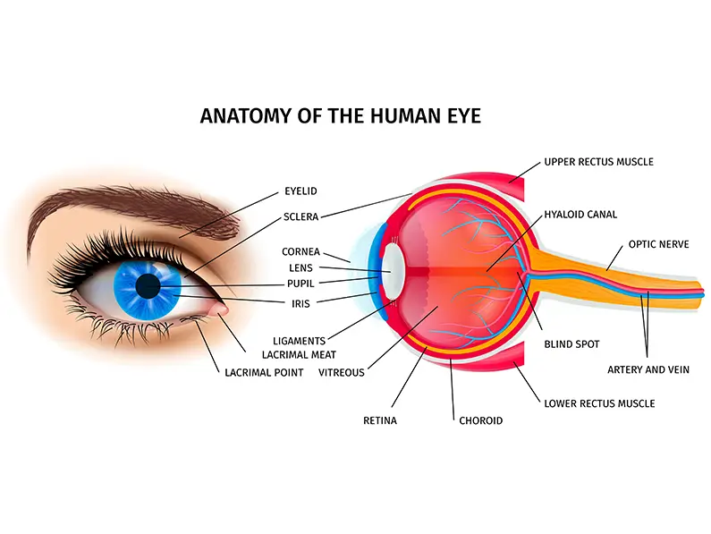
When we talk about eye anatomy, we’re referring to the structure and components that come together to enable vision. From the outer protective layers to the intricate inner parts, every segment of the eye has a purpose. By understanding these parts, you not only learn how vision works but also why maintaining eye health is so important.
Why Understanding Your Eye Matters
Think about the last time you marveled at a beautiful sunset or read a compelling book. Your eyes made that experience possible! Recognizing how these tiny structures function can encourage you to take better care of them. It’s a bit like understanding the engine of your car—when you know what each part does, you’re more likely to maintain it and prevent problems before they occur.
The Role of Each Part in Vision
Each element in your eye plays a unique role. Some parts protect your eye, others focus light, and still others convert light into signals your brain can understand. This orchestration of functions is what makes the miracle of sight possible. Let’s break down these elements further, starting with the basic layers that form the backbone of the eye.
Layers of the Eye
Our eyes are structured in layers, each serving a distinct purpose. Understanding these layers is essential to appreciate how your eye remains resilient and functional throughout your life.
The Sclera: The Outer Protective Layer
The sclera is the white, opaque outer layer of the eye. Much like a sturdy shield, it protects the inner components from injury and infection. This tough layer also helps maintain the shape of the eye, ensuring that other structures like the retina and choroid remain properly aligned for optimal vision.
The Choroid: The Nutrient Supply Layer
Nestled between the sclera and the retina, the choroid is rich in blood vessels. This vascular layer plays a pivotal role by supplying oxygen and essential nutrients to the eye’s tissues, especially the retina. Think of the choroid as the unsung hero that keeps your eye nourished and functioning.
The Retina: The Light-Sensitive Layer
The retina is perhaps the most critical part when it comes to vision. It’s a thin layer of tissue that lines the back of the eye and is loaded with millions of photoreceptor cells. These cells convert incoming light into electrical signals that the brain interprets as images. Without the retina, even the best-focused image would remain a mystery to your brain.
Functions of Key Structures
Beyond the basic layers, there are specialized structures that fine-tune our vision. Let’s zoom in on the lens, iris, and cornea, each of which plays a specific role in how we see the world.
The Lens: Focus and Clarity
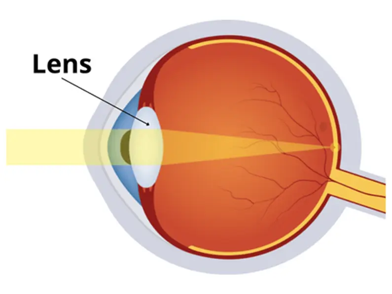
The lens of your eye is a transparent, flexible structure located right behind the iris. Its main job is to focus light onto the retina, a process that allows you to see objects clearly whether they’re near or far. This focusing ability, known as accommodation, is similar to adjusting the focus on a camera. As you age, the lens can become less flexible—a condition known as presbyopia—which is why many people need reading glasses later in life.
The Iris: Regulating Light
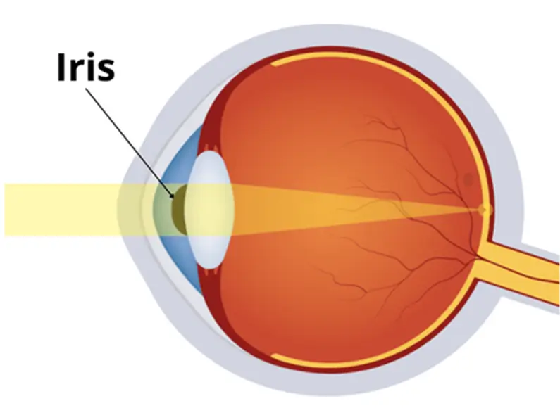
The iris is the colored part of your eye and works much like a camera’s aperture. It controls the amount of light that enters the eye by adjusting the size of the pupil. When you’re in a bright environment, the iris contracts to reduce the amount of light entering the eye, protecting the sensitive retina. In dim lighting, it dilates, allowing more light in to help you see better.
The Cornea: The First Line of Vision
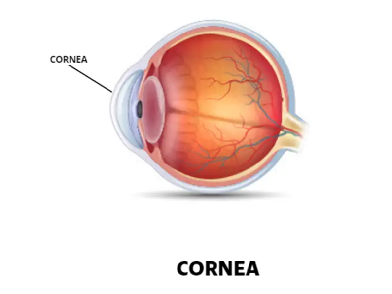
The cornea is the clear, dome-shaped surface that covers the front of the eye. Acting as the eye’s outermost lens, it plays a critical role in refracting (bending) light so that it properly reaches the lens and then the retina. Its smooth, transparent nature not only contributes to clear vision but also provides a protective barrier against dust, germs, and other environmental hazards.
Modern Imaging Technologies for the Eye
Advancements in imaging technologies have revolutionized the way we understand and treat eye conditions. These cutting-edge tools allow ophthalmologists to see the eye’s internal structures with remarkable detail, often before symptoms even arise.
Optical Coherence Tomography (OCT)
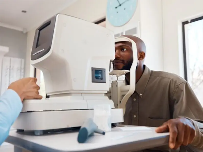
Optical Coherence Tomography, or OCT, is one of the most powerful imaging tools available today. It uses light waves to capture cross-sectional images of the retina, much like an ultrasound uses sound waves. OCT has become essential in diagnosing retinal diseases such as macular degeneration and diabetic retinopathy, enabling early intervention and better treatment outcomes.
Ultrasound Imaging and MRI in Ophthalmology
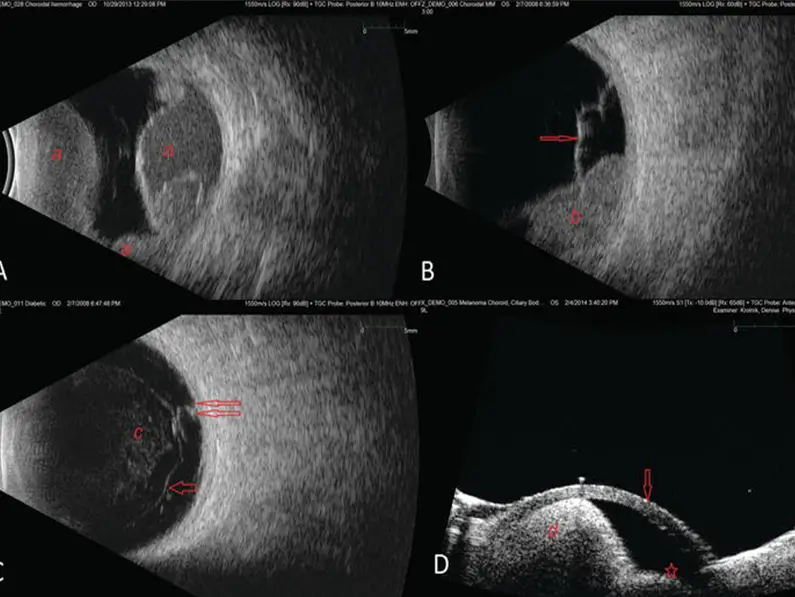
While OCT offers incredible detail of the retina, ultrasound imaging and MRI provide a broader view of the eye and surrounding structures. These imaging techniques are especially useful when examining deeper tissues or when other methods might be less effective. They help clinicians detect issues like tumors or structural abnormalities that might not be visible through standard examination methods.
Common Anatomical Variations in the Eye
Not all eyes are built exactly the same. Anatomical variations can occur naturally, and while many are harmless, some may influence how your vision works. Understanding these differences is important for both patients and eye care professionals.
Myopia, Hyperopia, and Astigmatism
Most of us are familiar with refractive errors like myopia (nearsightedness), hyperopia (farsightedness), and astigmatism. These conditions occur when the shape of the eye prevents light from focusing directly on the retina. Myopia is typically the result of an eye that is too long, while hyperopia occurs when the eye is too short. Astigmatism, on the other hand, is caused by an irregular curvature of the cornea or lens, resulting in blurred or distorted vision.
Rare Anomalies and Their Impact
Beyond common refractive errors, there are rarer anatomical variations that can affect vision. These might include congenital conditions, such as coloboma (a gap in one of the structures of the eye), or acquired changes due to injury or disease. Although these anomalies are less frequent, they can have significant implications for a person’s overall eye health and often require specialized care and treatment.
Implications for Eye Health
Understanding the anatomy of your eye is not just an academic exercise—it has real-world implications for maintaining optimal eye health. By knowing how each part functions, you can appreciate the importance of regular checkups, early diagnosis, and preventive care.
Preventative Care and Regular Checkups
Just as you’d take your car in for routine maintenance, your eyes also benefit from regular checkups. Early detection of issues like glaucoma, cataracts, or macular degeneration can make a huge difference in treatment outcomes. By keeping an eye on (pun intended) your vision and scheduling routine exams, you ensure that any potential problems are caught and managed early.
Impact on Daily Life and Future Trends
The structure of your eye affects more than just vision—it plays a role in your overall quality of life. From driving safely to enjoying a good book, clear vision is essential. Looking ahead, emerging research and technological advances promise even better ways to diagnose and treat eye conditions. Innovations such as artificial intelligence in imaging and minimally invasive surgical techniques are setting the stage for a future where eye care is more personalized and effective than ever before.
Conclusion and Takeaways
In summary, the human eye is a marvel of biological engineering. Its multiple layers and specialized structures work together seamlessly to capture and process the world around us. From the protective sclera to the nutrient-rich choroid and the light-sensitive retina, each part plays a crucial role in ensuring that we see clearly. The lens, iris, and cornea further refine this process, while modern imaging technologies have revolutionized the way we diagnose and treat eye conditions. Whether it’s common refractive errors or rare anomalies, understanding your eye’s anatomy is key to preserving its health.
Remember, your eyes are not just tools for sight—they are windows to your experiences, emotions, and memories. So, take the time to appreciate them, schedule regular checkups, and stay informed about the latest advancements in eye care. Here’s to seeing a bright, clear future!
FAQ: Your questions answered
What is the most critical part of the eye for vision?
The retina is arguably the most critical component because it contains the photoreceptor cells that convert light into electrical signals for the brain to interpret. Without a properly functioning retina, clear vision would not be possible.
How does the lens contribute to clear vision?
The lens adjusts its shape to focus light precisely onto the retina, allowing you to see objects at various distances. This process, known as accommodation, is vital for both near and far vision.
What role does the iris play in eye health?
The iris controls the size of the pupil, thereby regulating the amount of light that enters the eye. This adjustment not only aids in vision but also protects the inner eye structures from excessive light exposure.
How do modern imaging technologies improve eye care?
Technologies such as Optical Coherence Tomography (OCT), ultrasound imaging, and MRI allow clinicians to view the internal structures of the eye in great detail. This helps in the early diagnosis and treatment of various eye conditions, ensuring better outcomes.
Can common vision problems like myopia or astigmatism be corrected?
Yes, many common refractive errors, such as myopia, hyperopia, and astigmatism can be corrected using glasses, contact lenses, or refractive surgery. Regular eye examinations are crucial for determining the best corrective measures and maintaining optimal eye health.

